


July 18, 2021
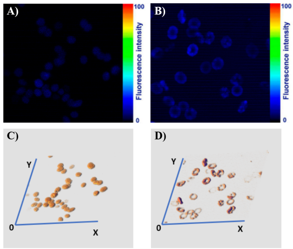
The research group headed by Dr. Gohar Tsakanova has published the results of the application of two-photon microscopy in the study of oxidative stress in human living cells in the Biological Journal of Armenia NAS RA (Tsakanova et al., “Qualitative and Quantitative Assessment of Oxidative Stress in Human Living Erythrocytes Using Two-Photon Microscopy Imaging Technique”, Biological Journal of Armenia, 2021, 1(73): 60-68). The study is carried out at DELTA two-photon microscopy station (CANDLE) by the scientists from the Institute of Molecular Biology NAS RA and CANDLE Synchrotron Research Institute.
Oxidative stress is connected to the enhancement of oxidizing agents’ production and a sharp decrease in the effectiveness of antioxidants. This can be a serious reason for the development of age-related human diseases, such as neurological diseases, pneumonia, cataract, cancer, diabetes, cardiovascular disorders, glaucoma, human aging, etc.
Red blood cells (RBCs) are one of the first cells exposed to endogenous and exogenous reactive oxygen species (ROS). At the same time, erythrocytes have strong antioxidant system, which allows keeping the oxidant/antioxidant balance in the organism. Nevertheless, various pathological states can lead to crucial changes in this antioxidant system and enzyme’s activity.
The main goal of present work is not only the analysis of RBCs using Two-photon microscopy imaging, but also the qualitative and quantitative assessment of oxidative stress, which will open new perspectives for the development of new and effective anti-aging substances. This new approach, used for estimating oxidative stress in human living RBCs, could successfully be applied in various fields of clinical research and analysis of antioxidant components.