


December 1, 2017
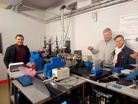
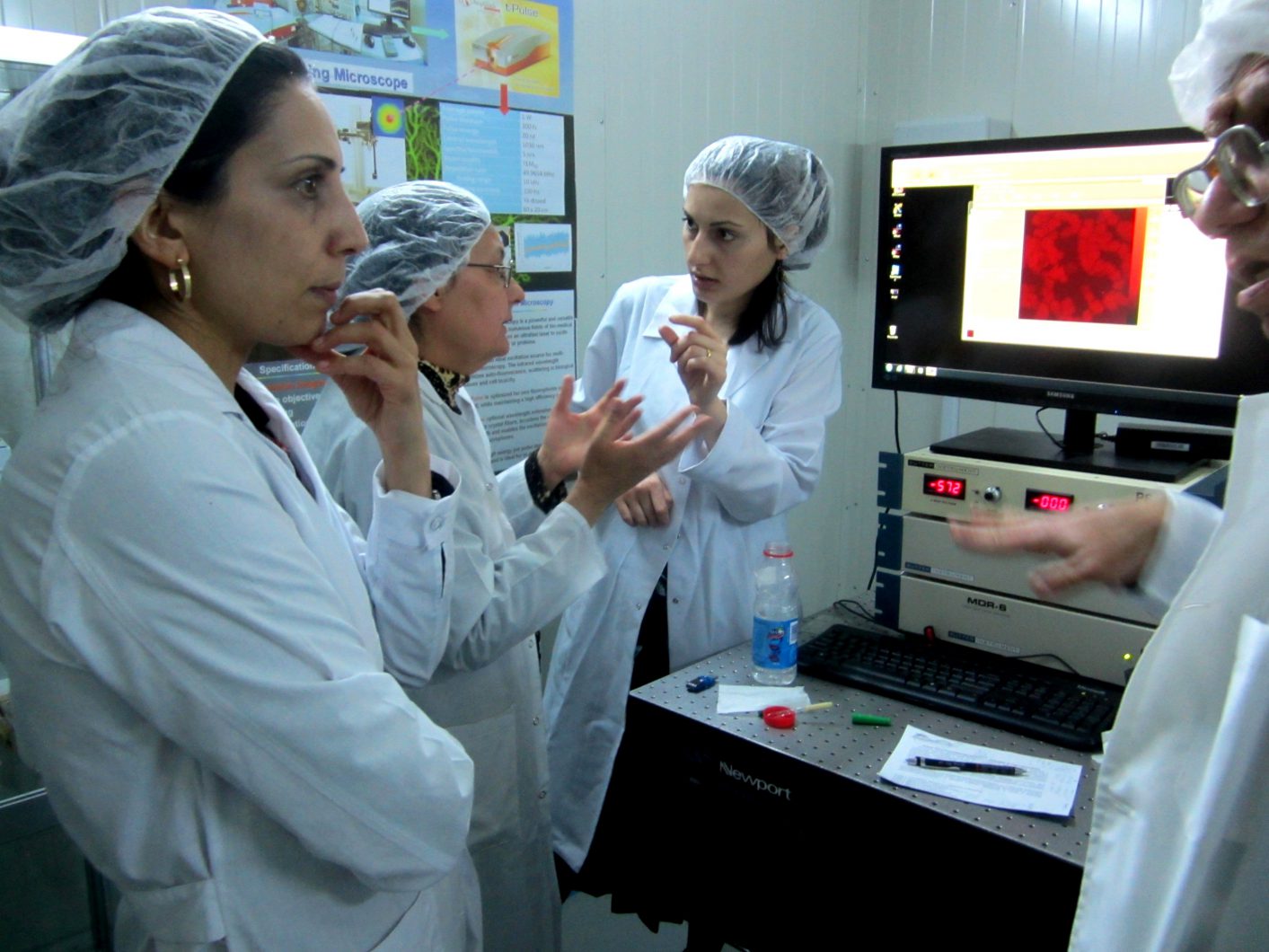
The article on “Two-photon microscopy imaging of oxidative stress in human living erythrocytes” has been published in the high-impact “Biomedical Research Express” journal of Optical Society of America (OSA) (https://doi.org/10.1364/BOE.8.005834, see also PDF variant). The work has been performed at DELTA two-photon microscopy station by the scientists from the Institute of Molecular Biology NAS RA, Yerevan State University and CANDLE Synchrotron Research Institute.
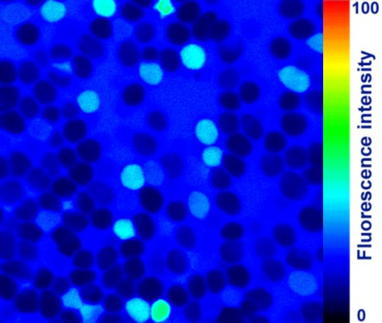
Red blood cells (RBCs) are known to be the most suitable cells to study oxidative stress, which is implicated in the etiopathology of many human diseases. The goal of the current study was to develop a new effective approach for assessing oxidative stress in human living RBCs using two-photon microscopy. To mimic oxidative stress in human living RBCs, an in vitro model was generated. The results of the oxidative stress were clearly visible on the two-photon microscopy images of RBCs as compared to no fluorescence in controls. This novel approach for oxidative stress investigation in human living RBCs could efficiently be applied in clinical research and antioxidant compounds testing.
The results open up new opportunities for real-time investigations of oxidative processes in human living cells that will contribute to a deeper understanding of the role of oxidative processes in pathomechanisms of various human diseases.